Ø Lower cost:
In upgrade:no need to obsolete original devices,cost only one third of purchasing new DR system.
In practical usage:film, developer solution and fixing solution are not necessary. The only cost is electricity fees.
Ø Operation time significantly reduced:
Old X-ray machine generally needs at least 5 hours from photographing to display due to processes of developing and fixing, etc. However, NAOMI captures digit image signals directly using the original X-ray projected to the detector through human body.Then digital image collection, processing, transition will be displayed on computor screen.The images could be directly used for diagnosis. It is only 6 seconds from photographing to display.
Ø Image definition upgraded to 48,000,000 pixel
There would be periphery deformation using CCD camera. NAOMI uses 192 high sensitive CCD camera arranged in matrix pattern to produce 48,000,000 pixel image
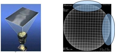
FIG 1.Periphery transformation by CCD camera of old DR machine
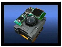
FIG 2. High sensitive camera
Non-effective deformation area |
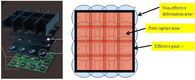
FIG 3. Advantageous spilt joint technology produced 48,000,000 pixel image and eliminates periphery deformation effectively.
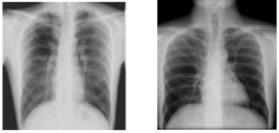
Film 96KV/6mAs NAOMI 96KV/0.375mAs
FIG 4. Clinical Case
Ø Sharply reduces maintenance cost
Normal DR applies amorphous selenium or single CCD. Once damaged, the whole develop board should be changed, which is extremely expensive. For NAOMI, only the CCD camera needs to be alternated if system damaged.
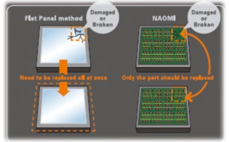
FIG 5. Only the CCD camera needs to be alternated
Ø Handy software system
Professionals could easily operate the system after calibration. The only thing you do is to open the power, click ‘take photo’. In 6 seconds, clear image will be displayed. |